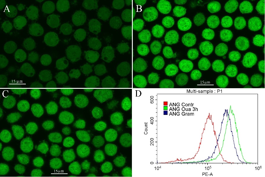
Fig. 2. Microscopy of ANG-stained U937 cells untreated (A), and treated with ouabain (B), or Gram (C). Cells were treated with ouabain for 3 h and stained with ANG-AM for the last 30 min, as described in the Methods section. Alternatively, cells were stained with ANG-AM in the presence of Gram in RPMI medium with serum for 30 min. After staining, cells were analyzed on a flow cytometer (D) or viewed on a confocal microscope (A-C).